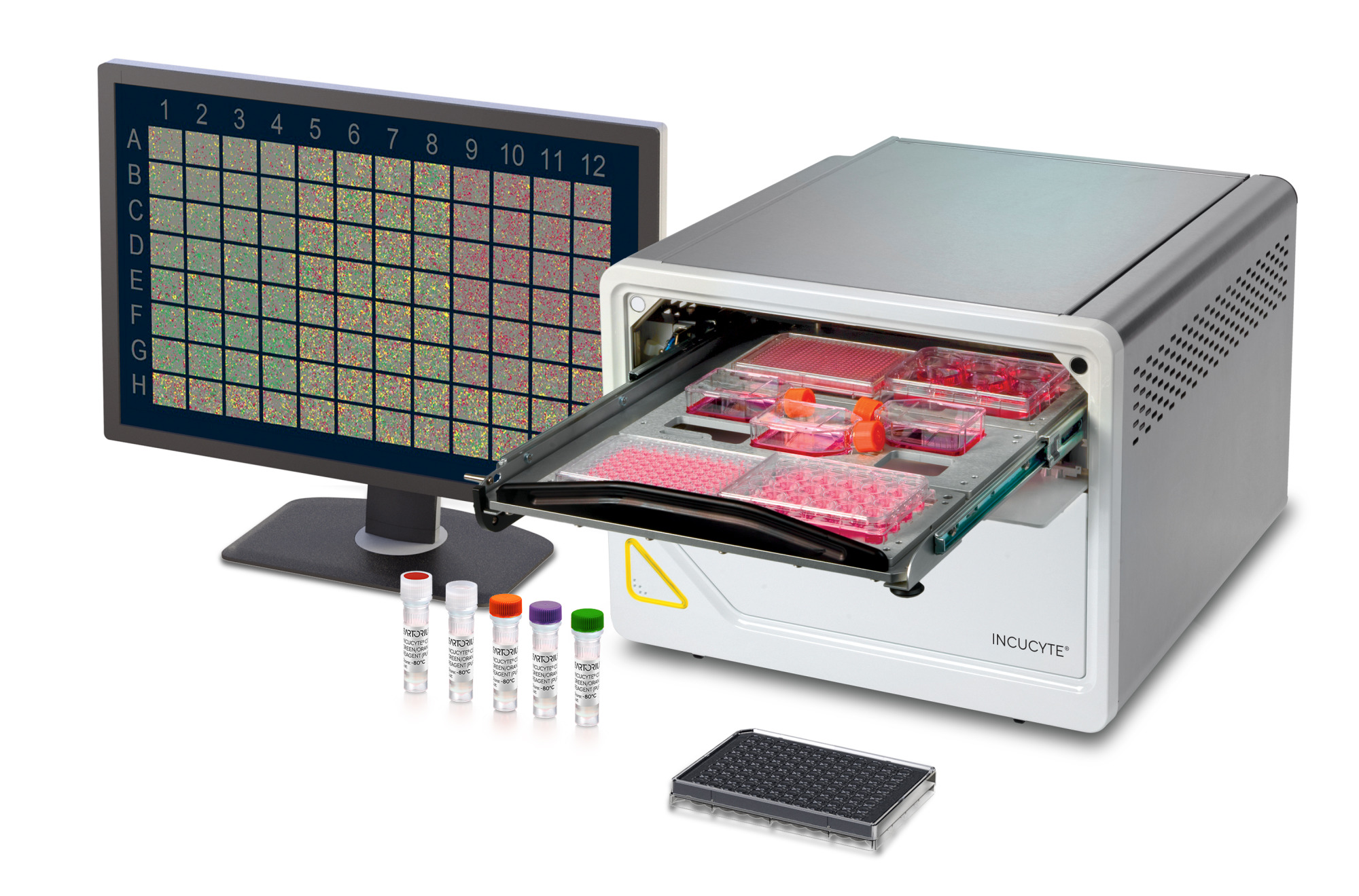
Live-Cell Imaging and Analysis Instrument: Incucyte® SX5
With the Incucyte® SX5 Live-Cell Analysis System, your research can flourish in a label-free, non-perturbing environment. See more information in every sample and explore more applications with patent-pending optics. Leverage up to five total fluorescence channels (up to three simultaneously) for long term time-lapse experiments, including a long wavelength, low phototoxicity NIR channel.
Key Features:
- Our most advanced application offering, including neuronal activity and metabolism
- Multi-user, multi-application support through remote network capability, unlimited free user licenses, independent vessel scheduling, and multi-objective scheduling.
- A Green/Orange/NIR optical module included, with optional Green/Red and Metabolism optical modules
- 4x, 10x, 20x objectives on an automated turret.
- Support for 3 interchangeable vessel trays and over 700 vessels, compatible with flask, dishes, slides, and microplates - up to 6 microplates in parallel.
Enjoy walk-away convenience as images are automatically acquired and analyzed in a variety of formats within the stable environment of a tissue culture incubator. Insight, accuracy and reliability is available at every step in the process, from culture maintenance to final experimental assay. Go where your research takes you with more colors, more insights, more possibilities!
Explore the Incucyte® SX5


See what the SX5 has to offer in Live-Cell Imaging & Analysis
More Colors. More Insights. More Possibilities.
Incucyte® SX5 Applications
Accelerate your next discovery with Incucyte®’s suite of live-cell applications. Gain real-time morphological and phenotypic insight for pathway and mechanistic studies by capturing time-dependent and cell-dependent treatment effects.
From the moment cells are placed in culture, the learning opportunity begins. Whether you’re working with rare or expensive cell types (e.g., primary cells, stem-cell derived cultures, patient derived cells) or recombinant cell lines, Incucyte® live-cell applications enable you to quantitate and validate cell morphology and phenotype during all stages of cell maintenance, modification as well as perform analysis on a wide range of phenotypic cell-based assays.
Make informed decisions about your cultures, rapidly optimize and improve your workflows, plus study complex live-cell assays to fast track your next discovery!
Accelerate your next discovery with Incucyte’s suite of live-cell applications.


Cell Monitoring & Workflows
Imaging Vessels
The Incucyte® Live-Cell Analysis Systems are compatible with a wider range of culture vessels – currently over 700 and growing! From 6-well to 384-well microtiter plates to standard tissue culture flasks, the Incucyte® can support your live-cell imaging and analysis needs.
SX5 Specifications
Category | Attributes | Description |
|---|---|---|
Optical System | Fluorescence Channels | Up to 5 |
Optical Module | Green/Orange/NIR (included) | |
Label-Free HD Phase Imaging | ||
Objectives | 4x, 10x, 20x (included) | |
Objective Automation | Automated Turret | |
Multi-User Support | Remote Network Capability | |
Incubation | Incubator Size | >200 L required |
Incubation to 42°C | ||
Software Module Compatibility | Incucyte® Cell-by-Cell | |
Capacity and Vesselware Compatibility | Interchangeable Trays | 3 |
Microplate Capacity | 6 | |
Microplate Compatibility | 6, 12, 24, 48, 96, 384-well | |
Flask Compatibility | T-25, T-50, T-75, T-100, T-175, T-225 | |
Other Labware Compatibility | 35mm, 60mm, 100mm, 150mm dishes and chamber slides | |
Computing and Storage | Operating System | 64-bit Windows 10 |
Controller Data Storage | 27.3 TB | |
Expandable RAID Storage | Add an additional 32.7 TB of storage with Incustore | |
Controller Memory | 64 GB |
Related Products


SX5 Resources
Featured Resources


FAQ for Live-Cell Imaging and Analysis Instrument: Incucyte® SX5
Incucyte® SX5 - Leverage up to five different fluorescence channels, up to three at a time (Green/Orange/Near-IR) plus HD Phase imaging, for long term time-lapse experiments. There is also the option of separately purchasing a Green/Red Optical Module or the Incucyte® SX5 Metabolism Optical Module, for use with the Incucyte® ATP Assay.
All models of the Incucyte® provide multi-user support with unlimited, free user licenses. Additionally, multiple assays can be run and imaged in different channels in parallel. However, only the Incucyte® SX5 and S3 support independent vessel scheduling for different image acquisition frequencies.
The Incucyte® itself is not an incubator but sits within a standard tissue culture incubator so cells are maintained in a physiologically relevant environment. For the Incucyte® SX5, an incubator larger than 200L is required.
The Incucyte® SX5 is our only instrument that, in addition to our phase channel, offers up to three fluorescent channels at one time with our Green/Orange/Near-IR Optical Module. This is a user swappable module that can be interchanged with the Incucyte® Green/Red Optical Module or the Incucyte® SX5 Metabolism Optical Module, for use with the Incucyte® ATP Assays. The Incucyte® SX5 also has an automated turret for the 4X/10X/20X objectives, as well as supporting independent scheduling of plates.
Because we want to provide the best and most relevant support to your research, Sartorius experts are available to demonstrate the features and benefits of our live-cell imaging and analysis systems in a personalized manner.
- Initial consultation with your local support team to understand your interests
- Virtual demo at no cost to you!
- Total demo time would be 2-4 hours, depending on number of applications
See how the Incucyte® can support your research needs, from setting up an assay to analyzing and exporting your data.